
Related items loading ...
Section 1: Publication
Publication Type
Thesis
Authorship
Temkov, Melissa
Title
Optofluidic Detection of Aqueous Ammonia and Parasitic Cysts
Year
2022
Publication Outlet
MacSphere Open Access Dissertations and Theses
DOI
ISBN
ISSN
Citation
Temkov, Melissa (2022) Optofluidic Detection of Aqueous Ammonia and Parasitic Cysts, MacSphere Open Access Dissertations and Theses,
http://hdl.handle.net/11375/30492
Abstract
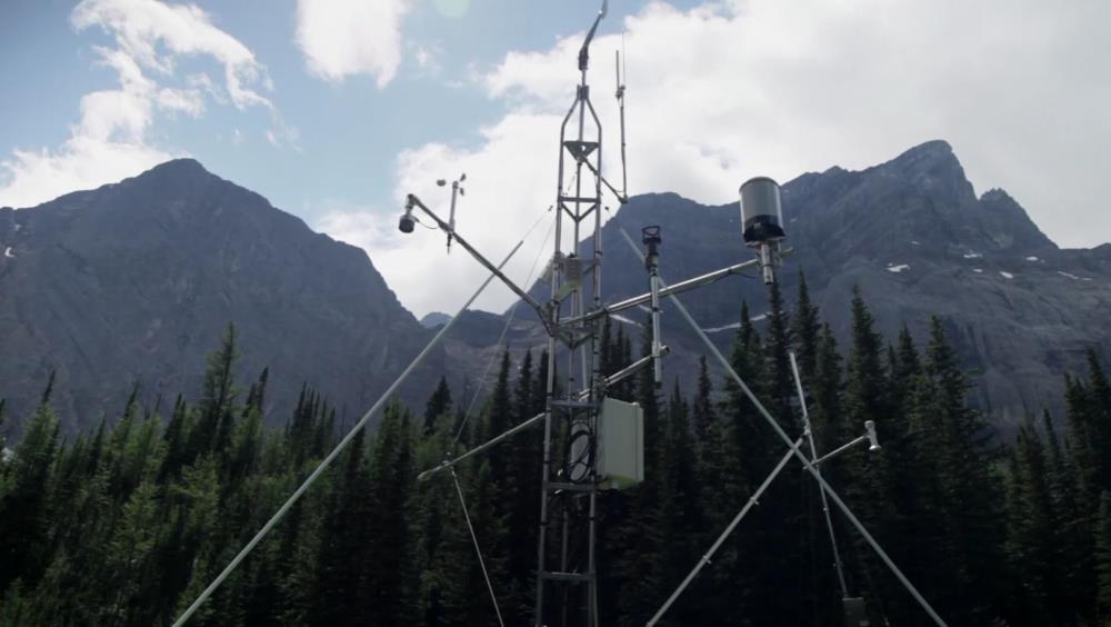
Water quality monitoring in Canada is essential to providing safe water to all. Indigenous and remote communities, many of which are under boiling drinking water advisories, lack availability and/or funding to water monitoring resources. A low-cost, point-of-care detection mechanism has been proposed for the detection of aqueous ammonia and protozoan parasites, which affect the safety of a source of water. An ammonia fluorescence responsive hydrogel, based on the fluorescence quenching of rare earth metal Europium (Eu3+) upon contact with aqueous ammonia, has been proposed to be incorporated into a microfluidic device, which utilizes shadow imaging and flow analysis to detect parasitic (oo)cysts of Cryptosporidium and Giardia, two of the most prevalent protozoan parasites which cause gastrointestinal illness around the world. Fabrication of the ammonia sensitive hydrogel was completed, and the essential components to the ammonia sensitivity were determined. Chemical analysis and solvent modifications found that Formamide is the essential solvent to maintain ammonia sensitivity. A literature review into the current detection mechanisms of Cryptosporidium and Giardia was completed to provide a reference and starting point for the development of the low-cost, point-of-care device proposed in this thesis. Baseline images of Cryptosporidium parvum and Giardia lamblia were captured to provide a reference for the development of a particle tracking algorithm to be used in the microfluidic device. The images captured highlight morphological features essential to developing a tracking mechanism based on the morphology of the (oo)cysts.
Plain Language Summary

 Metadata Editor
 Record List
 Alias List Editor
 Legacy sites

 GWFNet
 Master
 Research
 Map
Map
 Advanced
 . . .
. . .